


Granuloma annulare is a relatively rare, benign skin condition whose exact cause remains unknown. These lesions usually appear as small red or pink bumps that form a ring or semi-ring on various areas of the body, especially on the hands, feet, and sometimes the trunk. In many patients, this condition resolves spontaneously, but sometimes it is long-lasting and can even occur simultaneously with other immune disorders.
In a report published by Dr. Reza Moeini and colleagues in the Journal of Medical Case Reports , a rare case of the coexistence of granuloma annulare with alopecia areata and a type of dermatitis in a 6-year-old girl is described; a topic that could provide new insight into the overlap of skin diseases in children.
Clinical case description
The patient, a six-year-old girl from the Middle East, presented to the dermatology clinic with complaints of two skin lesions on the dorsum of the right foot, scaling of the soles and palms, pale patches on the cheeks, and patchy hair loss on the scalp. According to the mother, the first symptom appeared as eczema-like lesions on the soles of the feet, which spread to the palms and ventral surfaces of the fingers over a few days. Pale areas then appeared on the face, followed by patchy hair loss on the scalp. Finally, two distinct nodules developed on the dorsum of the right foot.
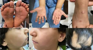
“A 6-year-old girl with (A) eczema, scaling, and clear lines on the soles of the feet; (B) red, itchy rashes on the palms of both hands; (C) red to pink nodules on the dorsum of the right foot, consistent with granuloma annulare; (D and E) pale patches on the cheek area with fine scaling, suggestive of pityriasis alba; and (F) patchy hair loss with a well-defined border.”
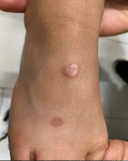
On examination, areas of alopecia were seen in the parietal region of the head with well-defined borders. On dermatoscopic examination, the characteristic feature of alopecia areata, “exclamation marks,” with black and yellow spots, was observed. There was no redness or scaling in these areas. The palms and soles had a red, itchy rash that in some places had developed into small, white-scaly ulcers, and the skin lines were more prominent. On the right leg, there were two well-defined red to pink nodules that were clinically consistent with granuloma annulare. On the face, there were symmetrical pale patches with fine scaling, consistent with pityriasis alba.
Dermoscopy findings
Close examination with a dermoscope revealed granuloma annulare lesions with fuzzy vascularity, a pink-red background, and yellow-orange areas without distinct structure. On the scalp, the classic signs of alopecia areata, including yellow and black spots and exclamation-mark hairs, were confirmed. On the palms and soles, a pink background with scattered punctate vascularity and white or yellow scaling were seen.
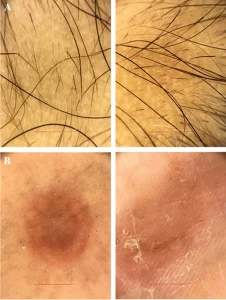
Dermoscopic images of granuloma annulare, alopecia areata, and palmar eczematous lesions in a 6-year-old girl; showing decentralised vessels, a pink-red background and structureless yellow-orange areas.
Treatment and follow-up process
To control lesions on the palms and soles, topical mometasone cream was prescribed twice daily for one week, along with a moisturizer and a mild cleanser, followed by calcineurin inhibitors. The nodules on the dorsum of the feet were also treated with the same steroid cream. For alopecia areata, topical minoxidil and betamethasone were used, and three intralesional injections of triamcinolone acetonide were given at one-month intervals.
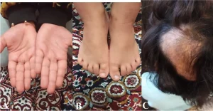
A) Palm lesions after treatment – B) Reduction of granuloma annulare – C) Remaining areas of alopecia.
After three months of follow-up, the palmar and sole lesions had completely resolved and the patient remained asymptomatic. The granuloma annulare lesions also responded well, but the patchy hair loss on the scalp did not resolve completely and some areas remained.
Discussion and analysis
This report is an example of the coexistence of multiple skin diseases in a child, a problem that is rarely addressed in clinical practice. The first symptoms in this child were eczematous lesions on the hands and feet, followed by pityriasis alba on the face. Alopecia areata appeared shortly after, and finally granuloma annulare nodules appeared on the feet.
Pityriasis alba is usually a mild manifestation of atopic dermatitis, but this patient did not meet the criteria for atopy (such as a history of allergies or asthma). Alopecia areata is a common autoimmune disease in children, with a peak incidence between the ages of two and six years, and can be associated with other immune diseases such as vitiligo or thyroid disorders. Granuloma annulare, although a benign and self-limiting lesion, has been associated with diabetes, thyroid disease, and dyslipidemia in some reports. The pathophysiology of GA is thought to be a delayed hypersensitivity reaction, but the exact cause of its co-occurrence with other immune disorders is still unclear.
In previous literature, the coexistence of GA and AA has been rarely reported and has often been considered a coincidental event. However, recording and publishing such cases could contribute to a better understanding of the shared immune pathways between these diseases and pave the way for future research.
Conclusion
Granuloma annulare is a benign condition that may occur alone or in association with other skin and immune problems. Paying attention to the pattern of lesions and considering concomitant diseases, especially in children, is of great importance in timely diagnosis and treatment. This report, published by Dr. Reza Moeini et al. in the Journal of Medical Case Reports and its translation is available on Dr. Reza Moeini’s blog , shows that the coexistence of granuloma annulare with alopecia areata and dermatitis, although rare, should be considered in clinical evaluation.
Source: Granuloma annulare with alopecia areata in a 6-year-old girl: a case report
To learn about a diverse collection of medical, health, and beauty articles, visit the Beauty and Health Articles section of Azargah Magazine .